This product is for research use only, not for human use. We do not sell to patients.
| Size | Price |
|---|---|
| 250mg | Get quote |
| 500mg | Get quote |
| 1g | Get quote |
Cat #: V42091 CAS #: 402615-91-2 Purity ≥ 99%
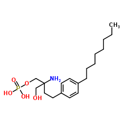
Description: Fingolimod phosphate (FTY-720-P) is the phosphate salt of Fingolimod (FTY-720; FTY 720; Gilenia and Gilenya) with improved water solubility. Fingolimod is an FDA approved drug for the treatment of Multiple sclerosis, acting as a S1P (sphingosine 1-phosphate) antagonist with potential antineoplastic activity. It inhibits S1P with an IC50 of 0.033 nM in K562 and NK cells. It is a folk medicine emerged from Fungi. Fingolimod was firstly found to be a therapeutic agent in organ transplantation. It plays the role in MS treatment through receptor-mediated actions both on the immune system and in the CNS. Fingolimod is used to treat multiple sclerosis, and has antineoplastic activity.
Publications Citing InvivoChem Products
Product Promise

- Physicochemical and Storage Information
- Protocol
- Related Biological Data
- Stock Solution Preparation
- Quality Control Documentation
| Molecular Formula | C19H34NO5P |
|---|---|
| CAS No. | 402615-91-2 |
| Protocol | In Vitro | In vitro activity: The inhibitory effect of S1P is revered by various concentrations of FTY720, with IC50 effect of 173 nM. In addition, FTY720 (10 nM) alone exerts no effect on the expression of co-stimulatory molecules. FTY720 reverses the increased expression of HLA-I induced by S1P for both the percentages of cells and the MFI, upon comparing the effect of S1P to the effect of combining S1P with FTY720. Medium and high-dose FTY720-P also enhances the levels of TGF-β1. TGF-β1 and Foxp3 mRNA expression are upregulated in the high-dose FTY720-P group. The proliferation of effector T cells is suppressed significantly in the medium and high-dose FTY720-P group at a Treg/Teff cell ratio of 1:1. At a ratio of 1:1, the proliferation of effector T cells is also suppressed in the high-dose FTY720 group. Kinase Assay: The monocyte-derived immature dendritic cells (iDCs) are pretreated with various concentrations of S1P for various periods of time prior to their incubation with NK cells. Four hours incubation of autologous or allogeneic iDCs with 0.2-20 μM of S1P significantly protectes these cells from NK cell lysis. The IC50 values of S1P are calculated at 160 nM for autologous iDCs, and 34 nM for allogeneic iDCs. Cell Assay: Immature DCs are left intact or are incubated with 2 μM S1P, 10 nM FTY720, 10 nM SEW2871 or the combinations of S1P with these drugs for 4 hours. As a control 1 μg/mL LPS is used. The cells are washed and incubated in a 96-well plate (v-bottom, 2 × 10⁵ cells per well), washed again and resuspended in PBS buffer containing 0.1% sodium azide. They are labeled with 1 μg/mL FITC-conjugated mouse anti-human CD80, 1 μg/mL FITC-conjugated mouse anti-human CD83, 1 μg/mL FITC-conjugated mouse anti-human CD86, 1 μg/mL FITC-conjugated mouse anti-human HLA-class I, 1 μg/mL FITC-conjugated mouse anti-human HLA-DR, 1 μg/mL FITC-conjugated mouse anti-human HLA-E, or 1 μg/mL FITC-conjugated mouse IgG as a control. The cells are washed twice, and examined in the flow cytometer. Markers are set according to the isotype control FITC-conjugated mouse IgG. To stain NK cells with antibodies for various NK cell activating receptors, they are either left untreated or incubated with 2 μM S1P for 4 hours, washed and stained with 1 μg/mL PE-conjugated mouse anti-human NKp30 (CD337), 1 μg/mL PE-conjugated mouse anti-human NKp44 (CD336), 1 μg/mL PE-conjugated mouse anti-human NKG2D (CD314), or as a control 1 μg/mL PE-conjugated mouse IgG1, for 45 min at 4 °C. NK cells are also stained with 1 μg/mL FITC-conjugated anti-killer inhibitory receptor (KIR)/CD158 antibody which recognizes KIR2DL2, KIR2DL3, KIR2DS2 and KIR2DS4, and as a control with FITC-conjugated mouse IgG. The cells are washed twice, and examined in the flow cytometer. Markers are set according to the isotype control PE-conjugated or FITC-conjugated mouse IgG. |
|---|---|---|
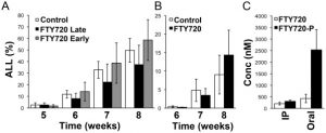
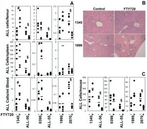
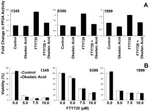
| In Vivo | FTY720 is effective in Ph+ but not Ph- ALL xenografts using an early disease model. FTY720 produces a significant reduction in disease burden in the Ph+ ALL xenografts using an early disease model. Ph+ human ALL xenografts responds to FTY720 with an 80 % reduction in overall disease if treatment has been initiated early on. In contrast, treatment of mice with FTY720 does not result in reduced leukemia compared to controls using four separate human Ph- ALL xenografts. | |
| Animal model | NOD/SCIDγc−/− mice bearing ALL cells. |
This equation is commonly abbreviated as: C1 V1 = C2 V2
- (1) Please be sure that the solution is clear before the addition of next solvent. Dissolution methods like vortex, ultrasound or warming and heat may be used to aid dissolving.
- (2) Be sure to add the solvent(s) in order.
